Last week, we had a particularly lively after-class discussion about dual-energy X-ray absorptiometry (DXA) scans and their possible pitfalls. Since this is one of the primary ways our bone health is diagnosed and monitored, it is important to understand the basics of the DXA examination.
DXA scans are not only used to help diagnose osteoporosis or low bone density (osteopenia), but also to help monitor patients who are receiving pharmacological treatment. However, there are instances when the test is not performed correctly which can lead to significant mistakes in diagnosis and therapy. As stated in one study, “A quality-control program must be implemented and followed to assure clinicians and patients that the information obtained falls within accepted ranges for precision and accuracy.” (Choplin et al., 2014).
Therefore, I am dedicating this month’s blog to a full review of the facts we should know about this test.
When and How Often to Repeat Your DXA Scan
The US Preventive Services Task Force recommends that all women 65 years and older be screened routinely for osteoporosis, or beginning at age 60 for women with additional risk factors. Men without risk factors should be screened beginning at age 70, or earlier in higher risk men. Some of these risk factors include prior fragility fracture, family history of osteoporosis or high fracture incidence, corticosteroid use, smoking, and excessive alcohol use.
After the initial test, it is typically repeated every two years, but may be up to five years, depending on the previous score as well as individual risk factors. If the first test did not show osteoporosis, waiting longer than two years will probably be recommended.
Which Body Parts Are Included and Why
Typically, DXA measurements of the lumbar spine (only L1 – L4) and one hip are performed because these are areas where serious, life-changing fractures can occur. The fifth lumbar vertebra is not included because of its proximity to the sacrum. And even though compression fractures of the thoracic spine are not uncommon, these areas are not scanned because the ribs, sternum (breastbone), lungs, heart, stomach, liver, etc., prevent a good view of this area.
If you have already had a fracture, especially in your spine, routine screening called Vertebral Fracture Assessment (VFA) is now being recommended by the International Osteoporosis Foundation. The picture below shows how this would be done.

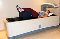
Fundamentals of Positioning
The lumbar spine is typically scanned first. Note the picture below showing the central line on the scanning table. The spine should be aligned as closely as possible to this line, and often the legs are positioned on a large foam block (as seen in the next picture), in order to bring the low back closer to the table. Elevating the legs has not been found to significantly affect accuracy, so it is not always done. The scan should include the lower portion of the 12th thoracic vertebra and the upper portion of the 5th lumbar vertebra. The interpreting radiologist will number each vertebra, and this will be used for careful comparison in subsequent testing.
To scan the hip, the leg must be straight and flat on the table. Positioning blocks are used to hold the leg in an inwardly turned position in order to get a better view of the hip joint. This angle should be between 15 and 25 degrees of internal rotation as can be seen in the third picture below.
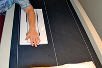
If one of the regions to be tested contains “artifacts” such as hip replacement hardware, pins, or screws, the forearm will be scanned instead. If degenerative disc disease of the spine or scoliosis are present, the forearm scan should be added to the overall test. Again, precise positioning is key to accuracy. Note the final picture.


T-scores and Z-scores
The T-score compares your bone density to that of a healthy 30 year old. A score of -2.5 or lower (more negative) is considered osteoporosis. A score of -1.0 to -2.5 is considered low bone density or osteopenia, and if your score is -1.0 or greater, your bone density is rated as normal. The Z-score compares you to a database of people in your own age range.
FRAX® Score
This calculation tool is now often included in routine DXA testing, and gives an estimate of your risk for fracturing a hip (or other major osteoporotic fracture) over the next ten years. You can use the link below to calculate this yourself. You’ll be prompted to answer simple questions such as your age, sex, weight, height, history of previous fracture anywhere, among a few others.
https://www.sheffield.ac.uk/FRAX
Calcium (Ca) Supplementation and DXA Scans
Undissolved Ca tablets in the gastrointestinal tract at the time of a DXA scan may affect the accuracy of the lumbar spine bone mineral density (BMD). In one study, an overlying Ca tablet had a substantial effect (12.6% increase) on the BMD of a single vertebral body (Kendler et al., 2006). Current guidelines are to avoid supplementation within 48 hours of testing.
Other Important Points
You will stay fully clothed, but make sure you wear comfortable clothing with no metal zippers, clasps or pocket coins. Naval jewelry should be removed.
Interference with accurate testing can also come from movement during scanning.
The test takes only about 10 minutes and the radiation emitted is one-tenth of a standard chest X-ray.
Quality Control
According to the International Society of Clinical Densitometry (ISCD), scanners should be tested by the facility for accuracy on a weekly basis, and a log of the results must be maintained.
Additionally, as noted by Choplin, et al. (2014):
“Because the output of the scanner depends on the training and diligence of the technician, each DXA facility should determine its precision error (PE) and calculate its least significant change (LSC).
The PE is the error expected in the test as a result of inherent machine fluctuations plus the imperfections due to the technologist’s positioning of the patient and selection of the regions of interest.”
The critical nature of this was highlighted in two other studies listed below. One found that out of 113 spine and hip reports, 61 hips and 94 spines were found to have images with incorrect positioning (Cetin et al., 2008). In the other, it was concluded that DXA technical and interpretation errors are “extremely common and likely adversely affect patient care” (Krueger et al., 2019).
For the most accurate results, it is recommended that you get tested on the same machine with the same technician. Otherwise, the following is recommended by the ISCD (listed by most to least preferred):
- same machine, different technician
- different machine within the same enterprise (with machines cross calibrated), same technician
- different machine within the same enterprise (with machines cross calibrated), different technician
Summary of the Limitations of DXA scans
- BMD is only one part of the story of fracture risk.
- The vast majority of fragility fractures, (fractures occurring after minimal trauma), occur in people who are NOT in the osteoporosis range (Cetin et al., 2008).
- Bone quality is not always measured, (unless trabecular bone score software has been acquired).
- Bone strength = BMD + bone quality
- Precision errors, quality control issues are not uncommon.
References
Cetin A, et al. Evaluation of the Patient Positioning During DXA Measurements in Daily Clinical Practice. Clinical Rheumatology 2008; 27:713-715.
Choplin RH, et al. A Practical Approach to Interpretation of Dual-Energy X-ray Absorptiometry (DXA) for Assessment of Bone Density. Current Radiology Reports 2014; 2:48.
Kendler DL, et al. Effect of Calcium Tablets on Interpretation of Lumbar Spine DXA Scans. Journal of Clinical Densitometry 2006 Jan-Mar;9(1):97-104.
Krueger D, et al. DXA Errors Are Common and Reduced by Use of a Reporting Template. Journal of Clinical Densitometry: Assessment and management of Musculoskeletal Health 2019; 22(1): 115-124.
Stone KL, et al. BMD at Multiple Sites and Risk of Fracture of Multiple Types: Long-term Results from the Study of Osteoporotic Fractures. Journal of Bone and Mineral Research 2003 November; 18(11): 1947-1954.